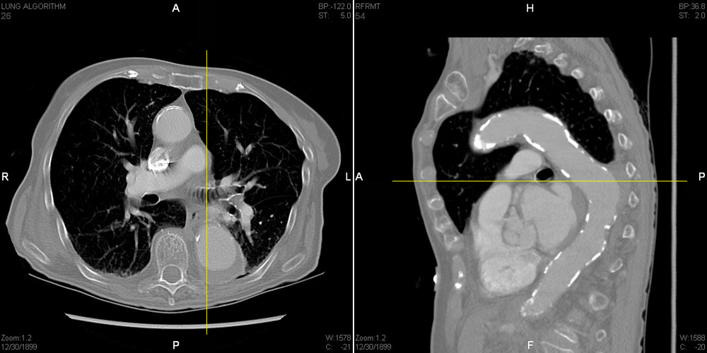
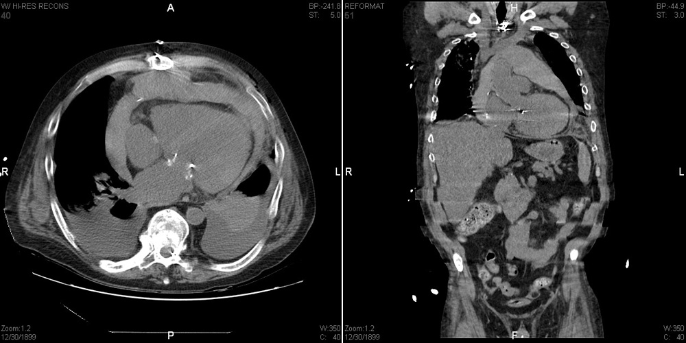
This is an example of a persistent left SVC. It was an incidental finding made by one of Stony Brook's very astute radiologists, Dr. William Moore. When I saw his write-up, I had to refer to an embryology text to be reminded not only that such a thing can occur, but how it happens. I will explain how a persistent left SVC arises, but it will be helpful if you also refer to the accompanying illustration, seen by clicking on the link below this text box.
Normally, a large shunt develops betweeen the left and right anterior cardinal veins. This shunt is the left brachiocephalic vein, which as you know carries blood from the left internal jugular and subclavian vv. to
the right side. After the shunt develops, the left common cardinal vein becomes deprived of most of its blood flow (the l. posterior cardinal v. having previously degenerated) and usually becomes fibrous. The part of the l. anterior cardinal vein inferior to the shunt persists as the l. superior intercostal vein, now carrying blood upward to the l. brachiocephalic vein. The part of the r. anterior cardinal v. inferior to the shunt, along with the r. common cardinal v., become the normal right-sided SVC. It empties into the smooth-walled part of the right atrium, derived from the right horn of the sinus venosus. The left horn of the sinus venosus becomes the coronary sinus. How then can one get a second SVC on the left side? It happens if the shunt between anterior cardinal veins fails to develop. As a result, the inferior part of the l. anterior cardinal v. and the l. common cardinal v. both stay big because they are carrying all the blood from the l. internal jugular and subclavian veins. Since the l. common cardinal vein empties into the l. horn of the sinus venosus, which has become the coronary sinus, the latter too is also much larger than normal. The full length of the l. anterior cardinal v., along with the l. common cardinal v. into which it empties, are said to constitute a left SVC. This left SVC in turn empties into the large coronary sinus. I don't expect anyone to remember this, but it was fun for me to review something I had long forgotten.
FOR iPhone USERS
This is an example of a persistent left SVC. It was an incidental finding made by one of Stony Brook's very astute radiologists, Dr. William Moore. When I saw his write-up, I had to refer to an embryology text to be reminded not only that such a thing can occur, but how it happens. I will explain how a persistent left SVC arises, but it will be helpful if you also refer to the accompanying illustration, seen by clicking on the link to the right of this text box.
Normally, a large shunt develops betweeen the left and right anterior cardinal veins. This shunt is the left brachiocephalic vein, which as you know carries blood from the left internal jugular and subclavian vv. to the right side. After the shunt develops, the left common cardinal vein becomes deprived of most of its blood flow (the l. posterior cardinal v. having previously degenerated) and usually becomes fibrous. The part of the l. anterior cardinal vein inferior to the shunt persists as the l. superior intercostal vein, now carrying blood upward to the l. brachio-cephalic vein. The part of the r. anterior cardinal v. inferior to the shunt, along with the r. common cardinal v., become the normal right-sided SVC. It empties into the smooth-walled part of the right atrium, derived from the right horn of the sinus venosus. The left horn of the sinus venosus becomes the coronary sinus. How then can one get a second SVC on the left side? It happens if the shunt between anterior cardinal veins fails to develop. As a result, the inferior part of the l. anterior cardinal v. and the l. common cardinal v. both stay big because they are carrying all the blood from the l. internal jugular and subclavian veins. Since the l. common cardinal vein empties into the l. horn of the sinus venosus, which has become the coronary sinus, the latter too is also much larger than normal. The full length of the l. anterior cardinal v., along with the l. common cardinal v. into which it empties, are said to constitute a left SVC. This left SVC in turn empties into the large coronary sinus. I don't expect anyone to remember this, but it was fun for me to review something I had long forgotten.

