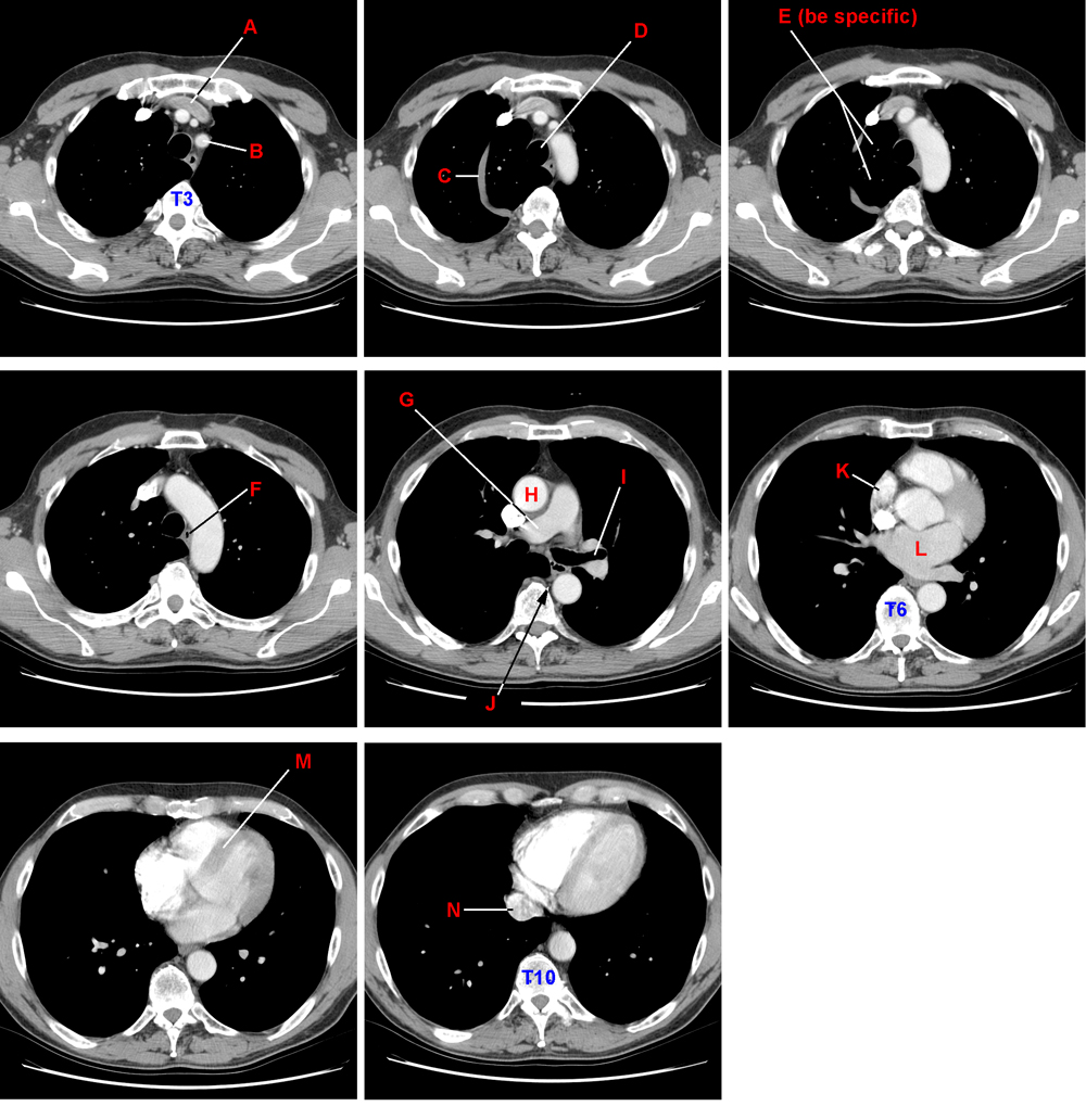
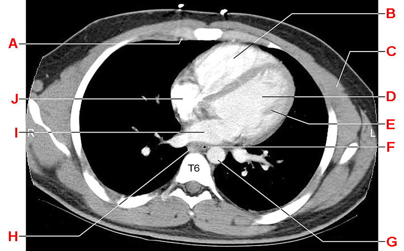
Presented below is a series of eight axial CTs of the chest. The most superior of the sections is at the upper left. The sections that follow are progressively more inferior. Contrast was injected into the right median cubital vein shortly prior to the imaging.
A =
B =
C =
D =
E =
F =
G =
H =
I =
J =
K =
L =
M =
N =
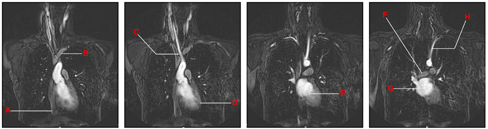
Presented below are a series of four MR angiograms at different (and sequential) coronal planes. The left image is the most anterior of the four; the right image is the most posterior of the four.
A =
B =
C =
D =
E =
F =
G =
H =
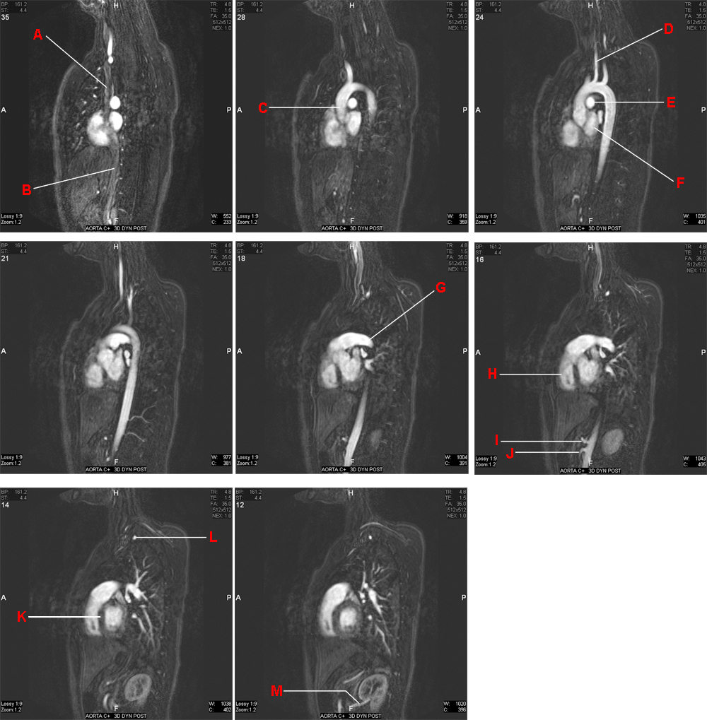
Presented below is a series of eight sagittal MR angiograms of the chest and upper abdomen. The rightmost of the sections is at the upper left (labeled 35). The sections that follow (labeled 28, 24, 21, 18, 16, 14, 12) are progressively further to the left.
A =
B =
C =
D =
E =
F =
G =
H =
I =
J =
K =
L =
M =
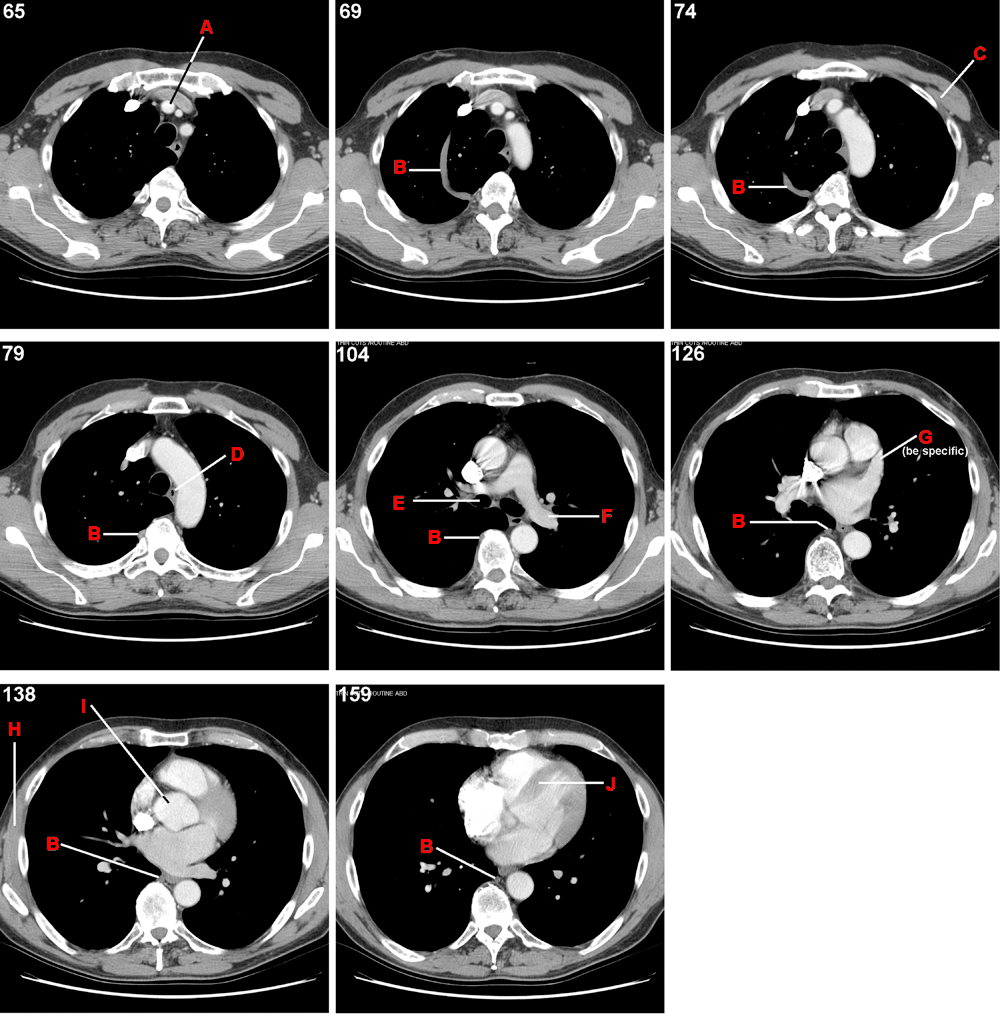
Presented below is a series of eight axial CTs of the chest. Each section is numbered in its upper left corner. Lower numbers are more superior. Each unit difference in this number corresponds to a distance of 1.25 mm. Thus, the last section is 11.75 cm below the first one. Contrast was injected into a vein of the right arm shortly before the images were taken.
A =
B =
C =
D =
E =
F =
G =
H =
I =
J =
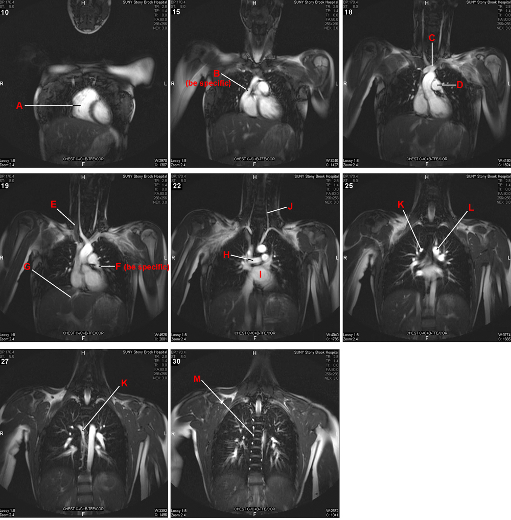
Presented below is a series of eight coronal MR angiograms of the chest. The most anterior of the sections is at the upper left. The sections that follow (indicated by higher numbers in the upper left corner) are progressively more posterior, but not by equal increments (a unit change in image number corresponds to 8 mm).
A =
B =
C =
D =
E =
F =
G =
H =
I =
J =
K =
L =
M =
Contrast was injected into this patient's r. median cubital v. immediately prior to taking the CT image presented below.